

History
Image 2.25
Wilhelm Rontagen observed and recorded x-rays when he experiemented with Hitterf-Crookes tubes. He observed that a piece of barium platinocyanide gave off light when the tube was in use. He theorized radiation traveled across the room and struck the unkown chemical and caused fluroscence. This developed his experiment ot capture x-ray photo's of a human hand.
1895
Through advances in technology and research, molecular and medical imaging can evolve. The evolution of molecular and medical imaging can help with the understanding of diseases and help prevent and detect diseases at an early stage.
THE FUTURE


Yasuyoshi Watanbe is the director of Riken. A center that develops key technologyies for breathroughs in the medical field.
2008

Image 2.41
With the help of Raymond Damadian through General Electrical Medical Systems they were able to develop new enhancements with the only upright MRI machine, that gave more accurate scannings of the neck and brain.
2001

Image 2.40
Professor Yasuyoshi Watanbe, along with professor Bangt Langstrom were able to produce more than 50 types of new labeled compounds. This lead to the new evaluation method of "In Vitro PET Method" which uses a segment of a living brain.
1993-1997

Image 2.39
William Edelstein, through the work of Richard Ernst, demonstrated imaging of the obyd. An image that could be acquired in approximately five minutes.
Image 2.38

1980
David Goldenberg used radiolabeled antibodies to image tumors in humans.
1978
Image 2.37

Image 2.36
1897

Raymond Damadian produced the first MRI scan of a human body. This discovery led to the research and evaluation of tomographic layers.
1977
Richard Ernst proposed magnetic resonance imaging using phase and frequency,Frouier Transform.
1975
Image 2.35

Image 2.34
CT scan first invented by Godrey Hounsfield at EMI Laboratories. The CT scan provides dimensional optical images that can be manipulated and rotated for review by doctors.
1972

Raymond Damdian created images of rat tumors through the use of magnetic resonance imaging.
1971

Image 2.33
Paul Lauterbur pioneered the use of nuclear magnetic resonance (NMR). He devloped a technique now known as MRI, that involves the analysis of the data obtained to produce multi-dimensional images.
1967

Image 2.31 & 2.32
David Kuhl in the late 1960's succeeded in producing the worlds first tomographic image of the human body. This brought about realization of which probes invaluable in the early detection of cancers.
1962

Image 2.30
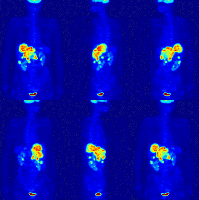
David Kuhl began experimenting in the late 1950's by taking cross-sectional images of distibution of radioisoptopes in the body which led to the development of CT, SPECT, and PET devices.
1954

Image 2.29

William H. Sweet helped advance the proton beam for brain tumors. While Gordon Brownell invented positron emission tomography which uses radioactive tracers to pinpoint the location of diseased tissue.
1953
Image 2.28

Image 2.26 & Image 2.27
Edward Purcell and Felix Bloch both contributed to nuclear magnetic resonance, without this NMR and MRI would not be discovered.
1946
Through the work of Henri Becquerel, Marie Curie was inspired and investigated the uranium rays. She then discovered two substances, Plochium and Radium, she found that these three substances are all "radioactive".
Henri Becquerel researched newly discovered x-rays. This led to the discovery of uranium rays.
Image 2.24
Text 1.13
Text 1.14,1.15
Text 1.16
Text 1.17
Text 1.18
Text 1.19
Text 1.20
Text 1.21
Text 1.22
Text 1.23
Image 2.42

History
Image 2.25
Wilhelm Rontagen observed and recorded x-rays when he experiemented with Hitterf-Crookes tubes. He observed that a piece of barium platinocyanide gave off light when the tube was in use. He theorized radiation traveled across the room and struck the unkown chemical and caused fluroscence. This developed his experiment ot capture x-ray photo's of a human hand.
1895
Through advances in technology and research, molecular and medical imaging can evolve. The evolution of molecular and medical imaging can help with the understanding of diseases and help prevent and detect diseases at an early stage.
THE FUTURE


Yasuyoshi Watanbe is the director of Riken. A center that develops key technologyies for breathroughs in the medical field.
2008

Image 2.41
With the help of Raymond Damadian through General Electrical Medical Systems they were able to develop new enhancements with the only upright MRI machine, that gave more accurate scannings of the neck and brain.
2001

Image 2.40
Professor Yasuyoshi Watanbe, along with professor Bangt Langstrom were able to produce more than 50 types of new labeled compounds. This lead to the new evaluation method of "In Vitro PET Method" which uses a segment of a living brain.
1993-1997

Image 2.39
William Edelstein, through the work of Richard Ernst, demonstrated imaging of the obyd. An image that could be acquired in approximately five minutes.
Image 2.38

1980
David Goldenberg used radiolabeled antibodies to image tumors in humans.
1978
Image 2.37

Image 2.36
1897

Raymond Damadian produced the first MRI scan of a human body. This discovery led to the research and evaluation of tomographic layers.
1977
Richard Ernst proposed magnetic resonance imaging using phase and frequency,Frouier Transform.
1975
Image 2.35

Image 2.34
CT scan first invented by Godrey Hounsfield at EMI Laboratories. The CT scan provides dimensional optical images that can be manipulated and rotated for review by doctors.
1972

Raymond Damdian created images of rat tumors through the use of magnetic resonance imaging.
1971

Image 2.33
Paul Lauterbur pioneered the use of nuclear magnetic resonance (NMR). He devloped a technique now known as MRI, that involves the analysis of the data obtained to produce multi-dimensional images.
1967

Image 2.31 & 2.32
David Kuhl in the late 1960's succeeded in producing the worlds first tomographic image of the human body. This brought about realization of which probes invaluable in the early detection of cancers.
1962

Image 2.30
David Kuhl began experimenting in the late 1950's by taking cross-sectional images of distibution of radioisoptopes in the body which led to the development of CT, SPECT, and PET devices.
1954

Image 2.29

William H. Sweet helped advance the proton beam for brain tumors. While Gordon Brownell invented positron emission tomography which uses radioactive tracers to pinpoint the location of diseased tissue.
1953
Image 2.28

Image 2.26 & Image 2.27
Edward Purcell and Felix Bloch both contributed to nuclear magnetic resonance, without this NMR and MRI would not be discovered.
1946
Through the work of Henri Becquerel, Marie Curie was inspired and investigated the uranium rays. She then discovered two substances, Plochium and Radium, she found that these three substances are all "radioactive".
Henri Becquerel researched newly discovered x-rays. This led to the discovery of uranium rays.
Image 2.24
Text 1.13
Text 1.14,1.15
Text 1.16
Text 1.17
Text 1.18
Text 1.19
Text 1.20
Text 1.21
Text 1.22
Text 1.23
Text 1.24
Text 1.29
Text 1.27, 1.28
Text 1.26
Text 1.25
Image 2.42